


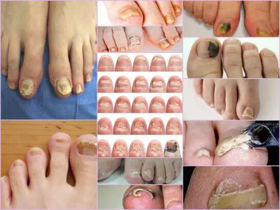
Onychomycosis is a type of fungal lesion, which exclusively affects the surface of the nails.To prevent the spread of timely infection, you need to know what the nail fungus looks like, as the therapeutic effect will be achieved faster if treatment began at an early stage of infection.Most often, the disease occurs in older people due to poor immunity, but the risk of getting a disease is about any person.There is no single classification of fungal nail damage;In medical practice, it is customary to distinguish them at the site of localization and the depth at which they penetrate.The infection is also grouped depending on the type of pathogen.
Types of Primary Pathogens and Signs
Symptoms of nail fungus in the initial stage are easily determined directly by the condition of the nail.This sign is more informative, as onycomycosis is always manifested in the form of changes in surface color, bed deformation, exfoliation and any external changes.The latter are expressed by rudeness, formation of grooves and cracks, as well as violation of cross and general nail points.
The main sign of a healthy nail is pink and transparency.Onychomycosis at each stage is characterized by the turbulence of the nail and the color change in yellow, brown or black green.At an advanced stage, the surface can get a black shade against the background of almost complete destruction.
External signs of infection with a fungus depend on the type of pathogen that infected.In medical practice, the following possible lesions are distinguished:
- Candida infection with a fungus, which is expressed by the discharge of the nail directly at the base of the box.Candidiasis of rollers around the nail will be characteristic of candidacy.This complication version may have either a bacterial source in the form of streptococci or staphylococci, or expressed in the form of medium plates or psoriasis;
- Dermatophytes of the Triachophyton section type.In this case, the penetration of the infection occurs directly from the free edge of the nail.The first symptoms of such a pathogen are the appearance of a yellow site on the surface.In the field of neoplasm, the nail is destroyed and the site itself tends to grow.The usual site of neoplasm localization is along the plate, parallel to the nail rolls, in this case, the infection is called distal-lateral.There is another form of loss from this pathogen - distal, in which the site appears in the part removed from the hole, mainly in the middle of the free edge;
Important!
This type of fungus most often occurs on the toe, gradually turning the nails into a loose yellow mass.In the absence of proper treatment, the disease is poured into hyperkeratosis.The nail plate is completely destroyed due to the spread of infection around the perimeter.
- Dermatophytes like trichophyton mentagophytes.Onychomycosis with such excitement is also most often based on finger nails, less frequently than little fingers.An infection with a fungus of this type requires not only the nail therapy but also the feet due to the rapid spread of the pathogen.A symptomatic disease can be like leikonichia, a common disease in medical practice.The main signs are white spots that appear in different parts of the nails, neoplasms are distinguished by a non -standard shape and different sizes.It is easy to distinguish fungi from leakonichia - in the latter case, the points are an air accumulation, which is not observed with fungal damage;
- Mold mushrooms.This possibility of damage is found significantly less frequently than a candidate or dermatophytic form.The main sign of such an infection - the surface of the plate acquires a dark, almost black shade.Not the whole finger can be completely infected, but only part of the nail plate.The first signs of nail fungus on the feet of this type are a sharp change of color.Onychomycosis can develop in the form of a longitudinal bar of black or dark green against the background of the rest of the pink part of the nail.
Diagnostics by type of change
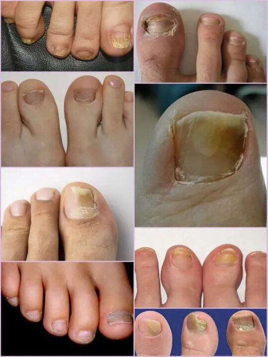
It is not difficult to notice a nail fungus in the leg even in the primary stages of infection, as an infection of this type manifests itself quite actively from the first day of the lesion.Instead of the usual transparent nails of pale pink in the patient, a significant surface deformation and a general change in condition are observed.The affected area has a dull yellow tinge, which appears mainly on the finger.The type of fungus and the degree of damage are the determining factors.
In the first stage, damage to the foot fungus standing is the appearance of small seals.The thickening of the plate and the keratinization of the bed under the nail will be characteristic.This stage is accompanied by a phenomenon such as partial detachment and discharge of the nail plate, which serves as a source of infection for healthy people.
Despite the active thickening of the plate, its constant grinding can be observed, despite the current factors.The characteristics of each stage and the symptoms of the fungus in the foot depend directly on the type of pathogen.
Depending on the changes that occur with the nail plate, three options are distinguished for onychomycosis:
- Hypertrophic;
- Normotrophic;
- Atrophic.
In the first case, there is a sharp change in the shade of the nail plate, its destruction along the edges, and the deformation of the surface of the plate.The nail thickens so much that it causes discomfort and painful sensations when walking.Normotrophic mycosis of the nails in the legs is distinguished by the presence of a healthy glow, but the plaque itself acquires some yellowness, stains on it.With an atrophic type of damage, brown and gray focuses on the nail surface.Thanks to them, it is possible to accurately determine the location of the pathogen.
Important!
The atrophic or onycolitical type of nail fungus is characterized by the thinning of the nail plate, and not its growth, as in other cases.Those areas in which the pathogen is localized tend to break away.Lack of proper treatment leads to an advanced stage - complete refusal of nail plate.
Classification of localization
Another sign by which you can divide the fungus damage to the fingers nails depends directly on the location of the focus on the nail plate.This also includes the depth of the pathogen, which in turn allows you to determine the approximate duration of future therapy and the chances of a rapid recovery.
Fungal Nail Diseases at the location of localization are classified into the following groups:
- Onychomycosis is a type of white surface - the appearance of many white spots on the surface of the nail plate.A fungus infection leads to the detachment of the skin at the places of the appearance of points where there is an active discharge of scales.The advanced stage leads to complete destruction and rejection of the plaque;
- Distal - develops at the free edge of the nail.The color change is first observed in a small area of the tile, after which there is an active expansion of its borders.The lesion is characterized by a yellow-gray or brown tinge, as well as an uneven rough surface and gradual exfoliation;
- Side atikomycosis has distal -like development stages, but is located exclusively on the side sides of the nail plate;
- Total infection - complete infection of the entire surface;
- Proximal onycomycosis.This disease begins its activity from the cuticle, which is inflamed, then the fungus quickly affect the nail, and the process itself begins with the appearance of a small white place near the inflamed area of the period.
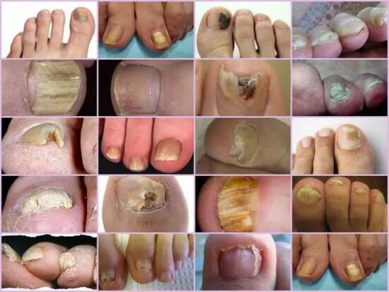
The most common forms of nail mycosis are side and distal, which is usually caused by dermatophytes.Such forms of damage as proximal and white can act as a secondary disease that accompanies the immune system's disease, for example, Vich.Total nail damage with a fungus should be considered as an advanced stage of development of each fungus under the nail.
Characteristics of fungi under the microscope
Despite the presence of an impressive number of onycomycosis signs, other diseases associated with skin problems and not the infectious nature in nature are accepted for fungal damage to the nails of the feet.You can determine the correct diagnosis and type of pathogen only in the laboratory under a microscope, for which, in a hospital, a piece of biological material from the affected areas is performed.
The resulting biomaterial is located in advance in an alkaline environment, after which a multiple increase is performed.Studying nail fungus under a microscope allows you to see an active pathogen, the outer form of which determines its type, the degree of distribution and an approximate degree of damage.In a nourishing medium, you can predict the approximate growth of the colonies, which allows not only to make an accurate diagnosis, but also determine the limitation of the infection.
Important!
Since it is possible to determine the presence of onycomycosis only in a laboratory, you should not postpone a visit to the doctor with the slightest suspicion.Nail fungi develop rapidly, and loss of time increases therapy.
What is an alarming signal?
Nail fungi in the legs are manifested by certain symptoms that are partly similar to some skin diseases.The following signs are more likely to indicate an onycomycotic lesion that requires medical intervention:
- The appearance of yellow spots on the plate, its deformation, a change in structure, which was not observed earlier;
- Darkening of the plate, loss of transparency, some photos of the nail fungus demonstrate a close shade of black, which is characteristic of pathogen forms;
- Obesity and keratinization of the nail plate, or vice versa very sharp;
- Rejection of the nail from the bed, the disconnection of scales and crumbs;
- Swelling of the roller, hanging it on the plate, the release of the fluid;
- Nails affected by the fungus appear inflamed, regardless of the type of pathogen.
All of these symptoms indicate a high risk of infection.Some external symptoms of changing the nail plate structure may be the result of other diseases.As fragility increases, the amount of calcium and iron in the body should increase, increased tuberosity means any infection of the body, with purely white stripes and dots, lack of copper or zinc is possible.Despite the fact that in the picture, nail fungus looks like a focal damage to the tile with a change in the color closer to yellow, this does not always indicate the infection.Yellowness can indicate various problems with the stomach and liver.
It is not difficult to recognize onycomycosis on the fingers from the external signs, despite the widespread classification of the disease.But you can determine the exact type of pathogen and the damage phase can only be in the laboratory.